
ELISA
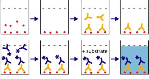
Step1 : Pre coat the Polysorp micro titer plate. Add 10µg of antigen/well in PBS and incubate for 1 hour at 37 °C . Flick out the contents and wash with PBS tween 20, 3 times. Again flick out the buffer and use the antigen coated plate immediately or wrap in an aluminium foil and store at -20°C until use.
Step 2: Block all the unbound sites to avoid false positives.
Step3 : Add antibody to the wells: Add 100µl of the antibody (starting at 1:50). Antibody is diluted in 5% goat serum in PBS buffer.Do a serial dilution. Add 200µl of antibody in the first well and 100µl of the plain PBS buffer in the subsequent wells. Then take 100µl of the antibody from the first well and add it to the second well. Mix it couple of times using the pipette and then take 100µl from the second well and add to the third well. Repeat the procedure. In the end discard the extra 100µl from the last well. Incubate for 30 minutes at 37 °C. Flick the contents into the sink and wash 3 times with PBS tween 20. Pat dry on a paper towel.
Step 4: Add anti-mouse IgG conjugated to an enzyme such as HRP (horse-radish peroxidase). Add 100µl/well diluted secondary antibody (1:5000). Incubate for 30 minutes at 37 °C. Wash with PBS tween 20, 3 times. Pat dry on a paper towel.
Step 5: Add 100 microliters of enhanced K blue TMB substrate to each of the wells. Incubate at room temperture for 30 minutes. You can see the blue color develop.
Step 6: Stop the reaction by adding 100µl /well 1N H2SO4. The blue color now turns yellow.Read at 450/650 nm using an ELISA reader.Result Interpretation: The intensity of the yellow colored developed is proportional to the amount of the enzyme linked antibody bound to the primary antibody which in turn is directly bound to the antigen. A standard curve is then constructed from which the antibody titer is determined.
The animal that produced the highest antibody titer will be used for bleeding and purifying the polyclonal antibody. However, if we are developing a monoclonal antibody against the antigen then we use the splenocytes from the mouse which has the highest titer to fuse with the myeloma cells for subsequent monoclonal antibody production.